Inheritance can be autosomal dominant, recessive or Xlinked. However, the most common types have an autosomal inheritance and are thought to be caused by mutations in the AMEL X gene, which codes for ameloblastin (C4), enamelin (C4) or tuftelin (Cl).
In the case of the autosomal dominant type of amelogenesis imperfecta, the locus of the defective gene is on chromosome 4q21 to which enamelin maps.
The less common X-linked types are caused by a variety of defects in the amelogenin genes and, confusingly, it seems the same mutation can sometimes cause hypoplastic, hypomineralisation or hypomaturation forms in different patients.
Genetic factors act throughout the whole duration of amelogenesis. Characteristically therefore, all teeth are affected and defects involve the whole, or are randomly distributed in the enamel. By contrast, exogenous factors affecting enamel formation (with the important exception of fluorosis) tend to
act for a relatively brief period and produce defects related to that period of enamel formation.
Hypoplastic amelogenesis imperfecta
The main defect is in formation of the matrix. The enamel is randomly pitted, grooved or very thin, but hard and translucent (Fig. 1). The defects tend to become stained, but the teeth are not especially susceptible to caries unless the enamel is scanty and easily damaged. |
| Figure 1. Amelogenesis imperfecta, hypoplastic pitted type |
The main patterns of inheritance are autosomal dominant and recessive, X-linked, and (a genetic rarity) an X-linked dominant type. In the last there is almost complete failure of enamel formation in affected males, while in females the enamel is ridged (Fig 2). Occasionally cases are difficult to classify (Fig. 3).
 | |||
| Figure 2. Close up of X-linked hypoplastic type amelogenesis imperfecta. These teeth from affected female show the typical vertical ridge pattern of normal and abnormal enamel as a result of lyonisation. |
 |
| Figure 3. Amelogenesis imperfecta, indeterminate type. Some cases such as this, are difficult to classify but are clearly inherited, as shown by their long family history. |
Hypomaturation amelogenesis imperfecta
The enamel is normal in form on eruption but opaque, white to brownish-yellow. The teeth appear similar to mottled fluoride effects (Fig 4). However, they are soft and vulnerable to attrition, though not as severely as the hypocalcified type.There are several variants of hypomaturation defects such as a more severe, autosomal dominant (type 4) of hypomaturation combined with hypoplasia.
 |
| Figure 4. Amelogenesis imperfecta, hypomaturation type. Tooth morphology is normal but there are opaque white and discoloured patches. |
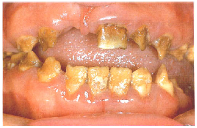
Hypocalcified amelogenesis imperfecta
Enamel matrix is formed in normal quantity but poorly calcified. When newly erupted, the enamel is normal in thickness and form, but weak and opaque or chalky in appearance.The teeth tend to become stained and relatively rapidly worn away. The upper incisors may acquire a shouldered form due to the chipping away of the thin, soft enamel of the incisal edge (Fig. 5). There are dominant and recessive patterns of inheritance.
 | |
| Figure 5. Amelogenesis imperfecta, hypocalcified type. The soft chalky enamel was virtually of normal thickness and form but has chipped away during mastication. |
Reference : Cawson, R. A. 2002. Cawson's Essential of Oral Pathology and Oral Medicine, vol. 7th ed. Churchill Livingstone, London.
No comments :
Post a Comment